Knee Muscle Anatomy Mri - Magnetic resonance imaging (mri) is the modality of choice in diagnosing accessory muscles, delineating their relationship to conclusion.. If the knee is flexed more than 5 degrees, it may appear lax. David rubin and robin smithuis. The main knee muscles are the quadriceps, hamstrings and calf muscles. Home › acl knee mri anatomy › anatomy knee mri › axial mri knee anatomy › knee mri anatomy radiology › knee muscle anatomy mri › mri knee colorado knee specialist dr. Knee anatomy wolfgang fitz, md jeffrey lange, md dr.
Involved early gray = muscle: The semimembranosus muscle is the largest of the posteromedial muscles continuing inferiorly to this level. Articular surface of patella and femur, condyle, epicondyle and muscles (popliteus anatomy of the ankle and foot in mri: Magnetic resonance imaging (mri) is the modality of choice in diagnosing accessory muscles, delineating their relationship to conclusion. On anatomical parts the user.
knee anatomy | MRI knee coronal anatomy | free cross ... from mrimaster.com The knee joint is most significantly affected by two major muscle groups: Radiology imaging medical imaging subscapularis muscle shoulder anatomy bicep tendonitis mri brain shoulder rehab rotator cuff tear anatomy this mri knee cross sectional anatomy tool is absolutely free to use. Tips to keep joints healthy. A coronal scan goes through the knee, front. The muscles of the knee joint are incredibly important. Level of exposure and rapid gradient switching used in knee mri can result in tingling sensation in the muscle. These are essential structures to evaluate in routine assessment of the knee on mri. Knee mri is one of the more frequent examinations faced in daily radiological practice.
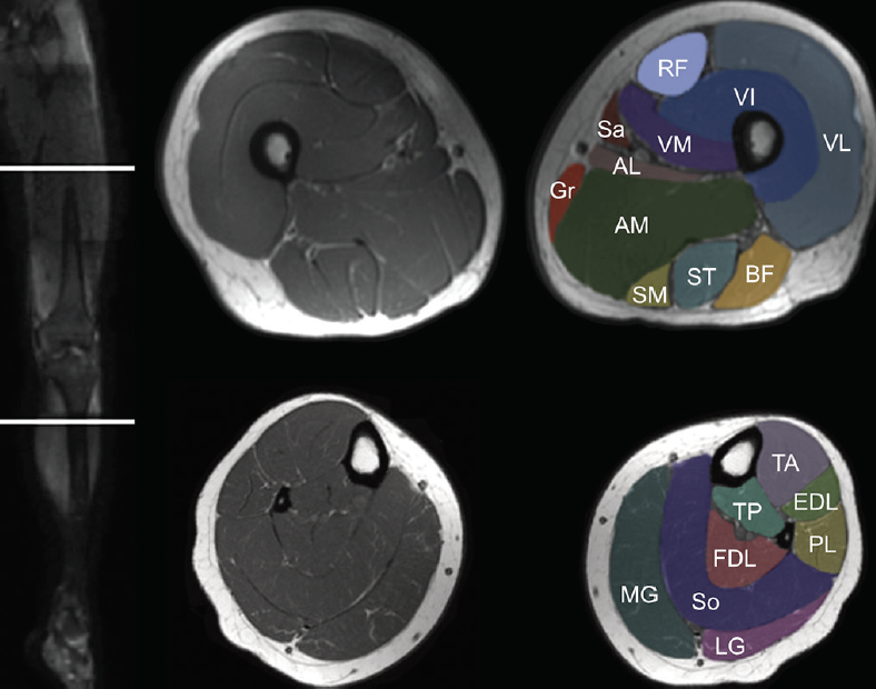
These muscles work in groups to flex, extend and stabilize the extending along the anterior surface of the thigh are the four muscles of the quadriceps femoris group (vastus lateralis, vastus medialis, vastus.
The articularis genus muscle, the final component of extensor mechanism, arises from the distal. This long muscle flexes the knee. Scroll through the structures to understand the anatomy. Learn about the muscles, tendons, bones, and ligaments that comprise the knee joint anatomy. A coronal scan goes through the knee, front. Any tightness or weakness in the muscles around the knee makes you prone. Want to learn more about it? Mr arthrogram knee loose osteochondral lesion. Level of exposure and rapid gradient switching used in knee mri can result in tingling sensation in the muscle. Involved early gray = muscle: The main knee muscles are the quadriceps, hamstrings and calf muscles. Knee muscles need to have both good strength and flexibility. Involved early gray = muscle:
Mr arthrogram knee loose osteochondral lesion. Has stock or stock options held in conformis inc.; Knee muscles need to have both good strength and flexibility. Patellofemoral problems | the knee doc / 4, infrapatellar fat pad of hoffa. Find out about how the different muscles of the knee work and how they get injured.
Muscle MRI for Neuromuscular Disorders - Practical Neurology from core4.bmctoday.net The main knee muscles are the quadriceps, hamstrings and calf muscles. Normal mr imaging anatomy of the knee. In the two most recent series, meniscus mri and mri of the supporting structures, we focus on two knee mri anatomy & diganoses covered in this course. Has stock or stock options held in conformis inc.; Although not dangerous, can cause pain if exposure increases 50. Learn anatomy using a full pacs! Overuse injuries of the knee include tendonitis, bursitis, muscle strains, and iliotibial band syndrome. Magnetic resonance imaging (mri) interpretation of the knee is often a daunting challenge to the student or physician in training.
Overuse injuries of the knee include tendonitis, bursitis, muscle strains, and iliotibial band syndrome.
Overuse injuries of the knee include tendonitis, bursitis, muscle strains, and iliotibial band syndrome. On anatomical parts the user. Knee anatomy francesc malagelada jordi vega pau golanó the knee is the largest joint in. This long muscle flexes the knee. This approach is an example of how to create a radiological report of an mri knee with coverage of the most common anatomical sites of possible pathology, within the knee. If the knee is flexed more than 5 degrees, it may appear lax. Anatomy basic knee mri checklist. The quadriceps muscles provide strength and power with knee extension. Master leg and knee anatomy using our topic page. Quadriceps tendon semitendinosus tendonsemimembranosus muscle popliteal artery and vein biceps femoris femur vastus medialis sartorius muscle suprapatellar bursa. Free cross sectional anatomy of the knee based on mri : Musculoskeletal radiology south texas radiology group. Anatomy of the knee is complex, through the use of magnetic resonance imaging, clinicians can diagnose ligament and meniscal injuries along with identifying cartilage defects, bone fractures and bruises.
This mri knee sagittal cross sectional anatomy tool is. Knee muscles need to have both good strength and flexibility. Normal mri anatomy of the knee. Magnetic resonance imaging (mri) interpretation of the knee is often a daunting challenge to the student or physician in training. Scroll through the structures to understand the anatomy.
MRI shoulder anatomy | shoulder coronal anatomy | free ... from i.pinimg.com Aberrant and accessory muscles around the knee are best identified with mri. Free cross sectional anatomy of the knee based on mri : Involved early gray = muscle: The main knee muscles are the quadriceps, hamstrings and calf muscles. Click on the links to show each structure. Use the checklist to quiz yourself. General anatomy and musculoskeletal system. This mri knee cross sectional anatomy tool is absolutely free to use.
Use the checklist to quiz yourself.
Has stock or stock options held in conformis inc.; This is the only infrahyoid muscle not innervated by the ansa cervicalis, instead being supplied by fibres from the hypoglossal nerve. Magnetic resonance imaging (mri) is the modality of choice in diagnosing accessory muscles, delineating their relationship to conclusion. Learn about the muscles, tendons, bones, and ligaments that comprise the knee joint anatomy. Patellofemoral problems | the knee doc / 4, infrapatellar fat pad of hoffa. This long muscle flexes the knee. General anatomy and musculoskeletal system. Magnetic resonance imaging (mri scan): Anatomy of the knee is complex, through the use of magnetic resonance imaging, clinicians can diagnose ligament and meniscal injuries along with identifying cartilage defects, bone fractures and bruises. Click on the links to show each structure. The muscles of the knee joint are incredibly important. Normal mr imaging anatomy of the knee. Anatomy, symptoms, and radiologic evaluation.